Look at two images below: a 19th-century Japanese woodblock print of a cresting wave, and a liver biopsy stained for iron. They share a color – the same deep, almost luminous blue. As pathologists, we spend our days reading colors and patterns to uncover truth in tissue. But we rarely stop to ask where those colors come from. I'd like to follow this particular blue, from a failed experiment in an alchemist's lab in Berlin, through the art of three continents, all the way to the slide on our microscope stage.
An accident in Berlin
The story begins in 1706, in the workshop of a pigment maker named Johann Diesbach, who was trying to make red dye from cochineal insects. Finding himself short of alkali, he approached his neighbor – an alchemist called Conrad Dippel – for some potassium salts.
Dippel is best remembered today for a foul-smelling concoction known as "Dippel's animal oil," distilled from blood and bones and once believed to have all manner of medicinal virtues. Diesbach was not to know that Dippel's salts were contaminated with residues of that animal oil – which is to say, with iron from blood. When Diesbach mixed them with iron sulfate, expecting to precipitate a vivid red, he got something else entirely. The mixture turned a deep, intense blue.
By sheer chance, he had produced iron(III) hexacyanoferrate(II) – the first modern synthetic pigment. We now call it Prussian blue.
It is worth reflecting on the improbability of this discovery. Diesbach was not searching for a blue, did not know any of the chemistry involved, and almost certainly didn't understand what had happened. A contaminated reagent, borrowed from the wrong shelf, gave the world a color.
A pigment changes the world
News of the new alternative traveled fast. By 1709, it had already appeared in European painting – most notably in Pieter van der Werff's Entombment of Christ.
But although Diesbach made it, it was artist and scientist Johann Leonhard Frisch who understood what it meant. Until then, the only reliable deep blue pigment available to artists was ultramarine, ground from imported lapis lazuli and worth its weight in gold. A canvas with serious quantities of ultramarine was a wealthy patron's flex. Frisch recognized the value of a synthetic alternative that was stable, vivid, and cheap.
He formally described the pigment in 1710 in Miscellanea Berolinensia, the journal of the Royal Prussian Academy of Sciences, and began producing it commercially. Within a decade, the European pigment market had been quietly reshaped. By 1724, after the recipe was leaked to London, Prussian blue had become a global commodity.
It found its way into the Prussian military uniforms that gave it part of its name, and into evening gowns for the London opera. On canvas, Lemoyne used it in Hercules Clubbing Cacus, and Gainsborough's famous Blue Boy owes much of its drama to the pigment. For the first time, artists had access to a deep, stable blue without bankrupting themselves. It became, quite literally, the color of an age.
The wave that traveled
What I find most remarkable is what happened when Prussian blue reached Japan through Dutch trade in the early 19th century. Before its arrival, Japanese woodblock artists worked mostly with indigo – a botanical dye with limited depth and a tendency to fade with light exposure. Prussian blue changed everything. It enabled richer tonal range, deeper shadows, and far better stability, and it gave rise to a whole genre called “aizuri-e”, literally, "blue prints."
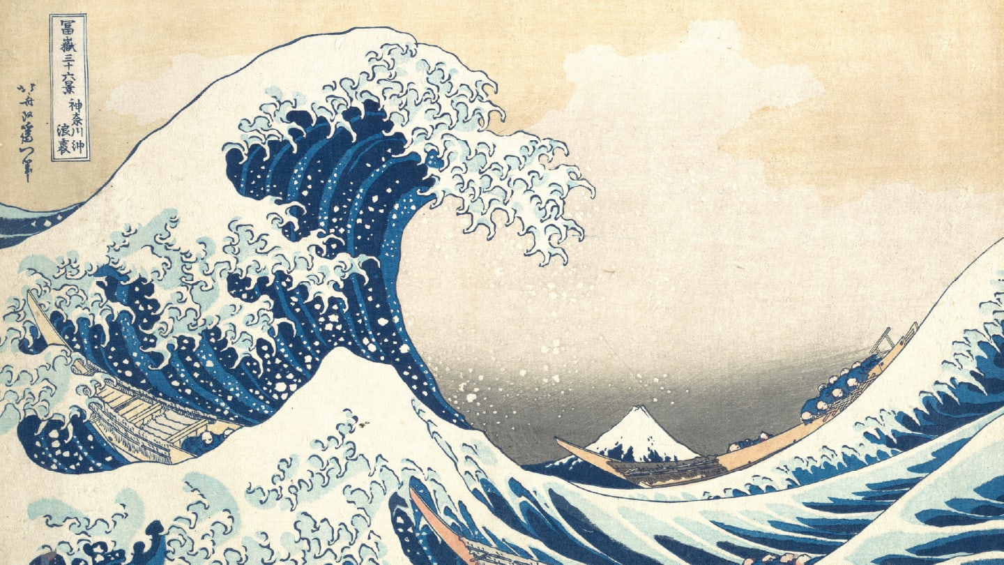
Around 1830, Katsushika Hokusai used the pigment in his Thirty-Six Views of Mount Fuji. The series includes the image I began this article with, Under the Wave off Kanagawa. A great wave crests over three fishing boats carrying fresh catch from the waters of Kanagawa toward the markets of Edo – today's Tokyo. In the distance, Mount Fuji stands small but steady, a quiet emblem of permanence against the chaos of the sea. The pigment that makes that wave possible, that gives the water its menace and depth, was of course, Prussian blue.
The prints then traveled back to Europe and helped shape the Japonisme movement of the late 19th century. You can see Prussian blue in the work of van Gogh, Picasso, and an entire generation of European artists. A pigment born from an accident in Berlin had gone around the world and changed art twice.
From canvas to cell
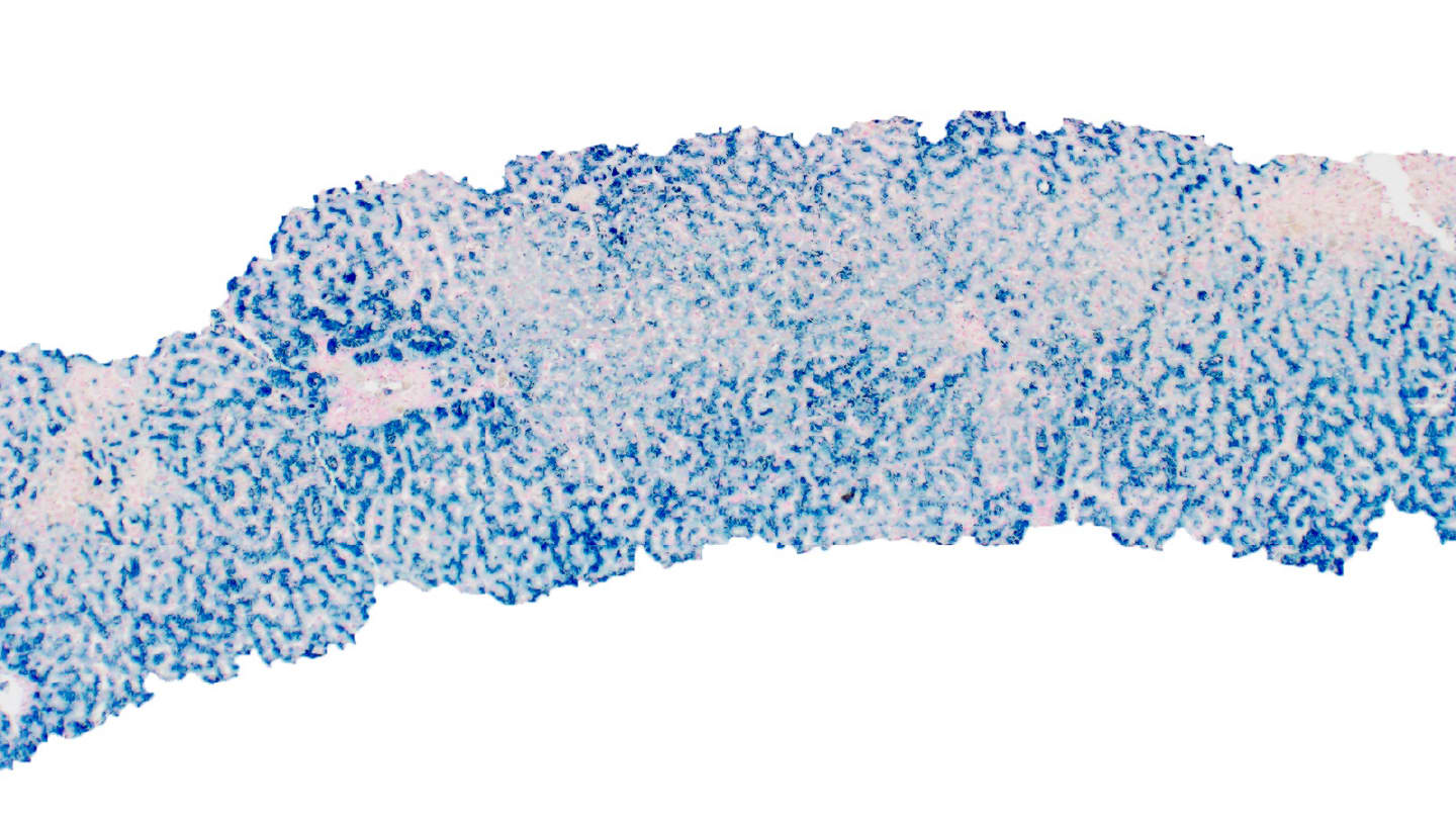
The pigment might have remained an art-history footnote if not for a young pathologist working at the Pathological Institute in Königsberg. In 1867, Max Perls was wrestling with a question that had been bothering pathologists for decades: those brown granules in tissue – were they melanin, or were they the remnants of degraded blood?
Perls took tissue from an old hemorrhage and treated it with potassium ferrocyanide in diluted hydrochloric acid. The acid liberated ferric iron from hemosiderin; the freed iron reacted with the ferrocyanide; and Prussian blue formed directly inside the cells where the iron lived. He repeated the experiment in lungs, spleen, and liver and showed that bile and fat stayed stubbornly unstained. With one elegant reaction, he had turned a pigment into evidence.
The chemistry hasn't changed since:
HCl releases Fe³⁺ from hemosiderin → Fe³⁺ + ferrocyanide → Prussian blue precipitate.
Heme iron, locked inside its porphyrin ring, isn't liberated by HCl, which is why the reaction is so specific for storage iron and not for the iron still bound in intact red cells.
Why we still reach for it
More than 150 years later, the Perls Prussian blue stain remains the gold standard for evaluating iron in liver pathology. It gives us both a semi-quantitative grade and, just as importantly, a qualitative read on where the iron sits, revealing the cell type, zone, and compartment: hepatocellular versus reticuloendothelial. The stain anchors the workup of hereditary hemochromatosis, transfusion-related iron overload, and the various chronic liver diseases in which iron quietly accumulates and contributes to fibrosis.
The reach extends well beyond the liver. In the lung, Prussian blue lights up the ferruginous bodies of asbestosis – iron-coated asbestos fibers with their unmistakable dumbbell ends, and the dust-laden macrophages of siderosilicotic nodules. In hematopathology, it's how we confirm ring sideroblasts in myelodysplastic syndromes: that telltale halo of iron-laden mitochondria encircling the nucleus of an erythroid precursor, requiring at least five granules covering a third of the nuclear circumference to count.
Forensic pathology has found its own uses for the stain. Hemosiderin-laden macrophages can help date hemorrhage in subdural hematomas, with iron typically becoming demonstrable around three days after bleeding. However, the reliability of precise dating remains debated, and overinterpretation is a known pitfall.
In hemorrhage, pairing the stain with glycophorin A immunohistochemistry can help distinguish an antemortem from a postmortem case. In button battery ingestions, metallic iron released into the surrounding mucosa becomes detectable as the tissue is injured. So-called "rust rings" or "rust stains" – iron transferred from a gun's trigger or barrel to the skin – can be demonstrated as supportive evidence of contact, because hydrochloric acid in sweat does much the same thing in vivo that Perls did in his beaker, releasing the iron to react.
An enduring legacy
In a final twist, Prussian blue is itself an FDA-approved therapeutic. Taken orally, the pigment binds radioactive cesium and thallium in the gut by ion exchange, prevents their reabsorption through enterohepatic circulation, and accelerates their excretion. The same molecule first synthesized by accident in a Berlin workshop is now stockpiled for use after radiological emergencies. The pigment has become an antidote.
Prussian blue was born in an alchemist's shadow, industrialized by chemists, taken up by artists from Berlin to Tokyo, and finally redeemed by a pathologist in Königsberg. Every time we drop a slide into Perls solution and watch the iron declare itself, we're using a reaction that ties our daily work to a chain of accidents, insights, and ambitions stretching back three centuries.
Teaser credit: Images for collage sourced from Adobe Stock
Newsletters
Receive the latest pathologist news, personalities, education, and career development – weekly to your inbox.

References
- M Perls, Archiv F Pathol Anat, 39 (1867). doi: 10.1007/BF01878983.
- AD Burt et al., "MacSween's Pathology of the Liver," 8th Edition (2022), Elsevier. doi: 10.1016/C2018-0-05272-X.
- ML Smith et al., "Practical Pulmonary Pathology," 4th Edition (2022), Elsevier.
- MM Patnaik and A Tefferi, Am J Hematol, 96, 3 (2021). PMID: 33428785.
- A Sonada et al., Forensic Sci Int Synergy, 11 (2025). PMID: 40686580.
- DR LaFrance et al., Forensic Sci Med Pathol, 7, 3 (2011). PMID: 21305390.


