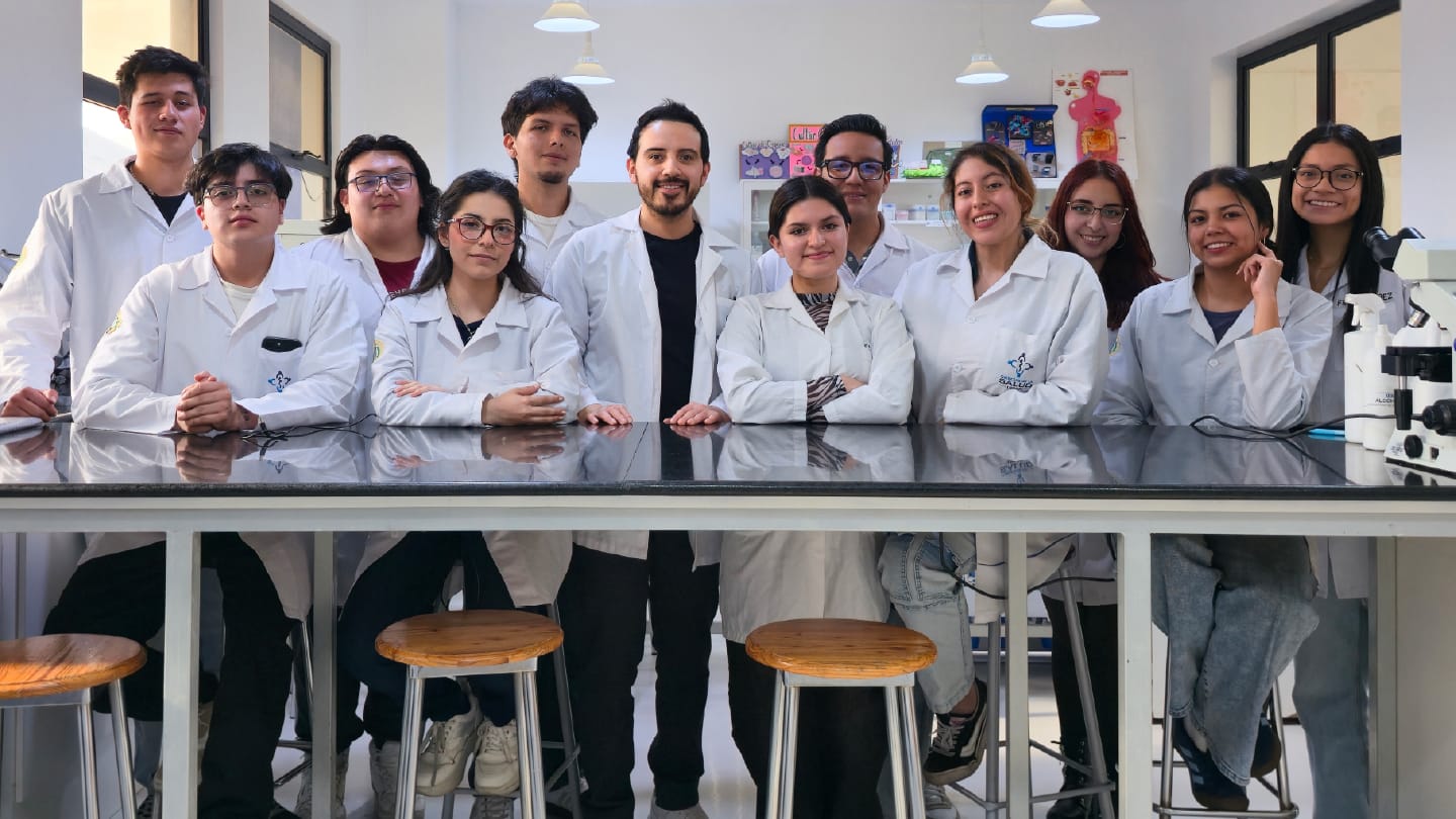
Class photograph (left to right): Alejandro Terán, Dylan Toctaquiza, Matheo Brito, Vallery Ruíz, José Aranda, Prof. Alex Zapata, Ariana Bastidas, Adrián Gallo, Milenka Guzmán, Camila Robayo, Ainhoa Mena, and Fiorella Páez.
Pathology education is not often associated with paint palettes, petri dishes, or classroom creativity. But Biomedical Scientist and Professor Alex Zapata is challenging that perception by bringing #PathArt into his teaching. Alongside examples of pathology-inspired artwork, Zapata invites students to create agar art of their own – a hands-on exercise that turns microbial growth into a visual learning experience.
In this article, Zapata opens the door to his classroom to explore how creativity can support microbiology education. We also hear directly from his students, who share their views on whether artistic approaches can make complex scientific concepts more engaging, memorable, and accessible.

What inspired you to start using #PathArt and other creative approaches in pathology and microbiology education?
Working in a tertiary-care hospital has taught me that pathology and microbiology require more than scientific knowledge. They demand careful observation, thoughtful interpretation, and the ability to connect laboratory findings with patient care.
Microbiological photography played a fundamental role in my learning. Capturing the macroscopic and microscopic features of microorganisms trained my eye to recognize subtle morphological details and appreciate the beauty and complexity of the microbial world. Agar art later offered a different perspective. By working with microorganisms in vitro, I gained a deeper understanding of how they grow, interact, and behave under laboratory conditions. Watching living organisms produce patterns, textures, and colors transformed how I viewed microbiology.
These experiences became the foundation of #PathArt. The initiative did not begin as an art project; it emerged at the Mycology and Tuberculosis Unit in the Hospital de Especialidades Carlos Andrade Marín, where photography and agar art naturally developed into educational tools. My goal was to bring that same spirit of observation, curiosity, and creativity into the classroom, helping students connect scientific concepts with meaningful visual experiences and clinical practice.
What visual tools or creative methods do you use in your teaching, and how do they help students understand complex concepts?
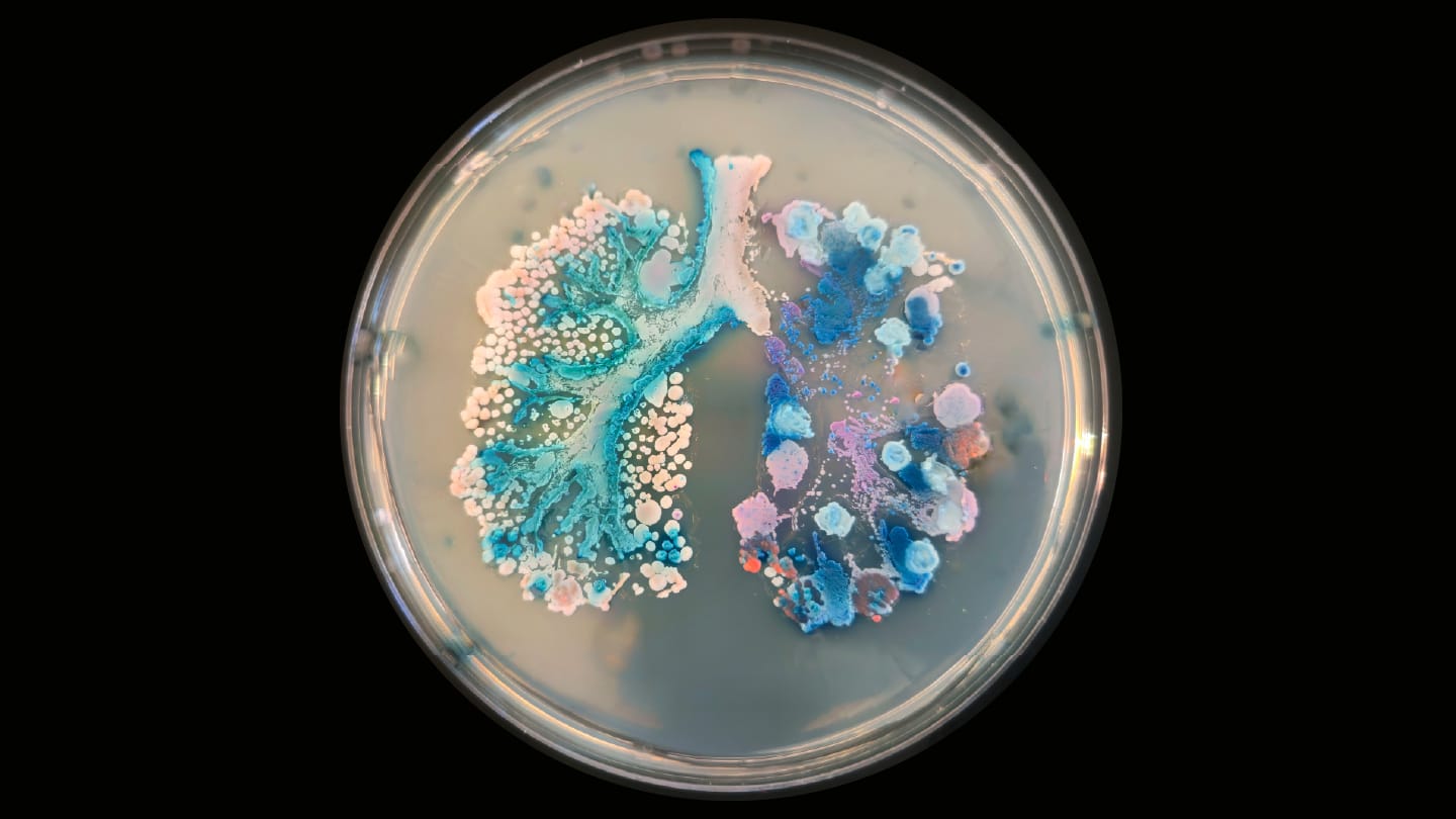
One of the main tools I use is agar art created from yeast colonies grown on chromogenic media. The natural pigmentation of different species allows students to produce anatomical, physiological, and microbiological illustrations while learning how chromogenic media support microbial identification.
I also incorporate microbiological photography into my teaching. High-quality images of colony morphology and microscopic structures allow students to compare conventional laboratory techniques with automated diagnostic methods. When paired with real clinical cases, these images help students understand the relationship between laboratory findings, disease, and patient care.
How can art help students develop skills that are important in pathology, laboratory medicine, and diagnostics?
Throughout history, art has helped people understand and communicate complex ideas. In pathology and laboratory medicine, it offers a unique way to transform abstract concepts into meaningful visual experiences. Colors become anatomical structures, textures represent tissues, and microorganisms form part of a broader clinical story. This approach strengthens observational skills, spatial awareness, creativity, and long-term retention, all of which are essential in diagnostic disciplines.
Can you share an example of a creative project or lesson that had a particularly strong impact on student learning?
One of the most rewarding experiences was organizing a #PathArt session in which students from Biomedical Sciences at Universidad Internacional used chromogenic yeast colonies to illustrate physiological processes, anatomical structures, and disease mechanisms. Rather than simply identifying microorganisms, they explored how colony colors and growth patterns could be transformed into scientific illustrations. This activity deepened their understanding of the microbiological principles underlying chromogenic media and the clinical significance of the diseases they depicted.
Ainhoa Mena
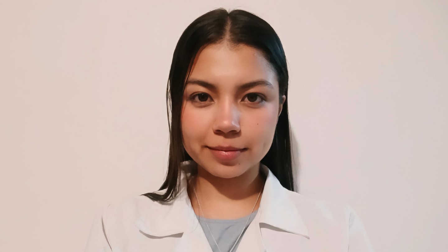

Agar art has deepened my understanding of microbial growth, colony morphology, culture conditions, and aseptic technique. It has also reinforced the importance of precision, biosafety, and contamination control. These activities encourage creativity while making scientific communication more engaging and accessible.
Vallery Ruiz
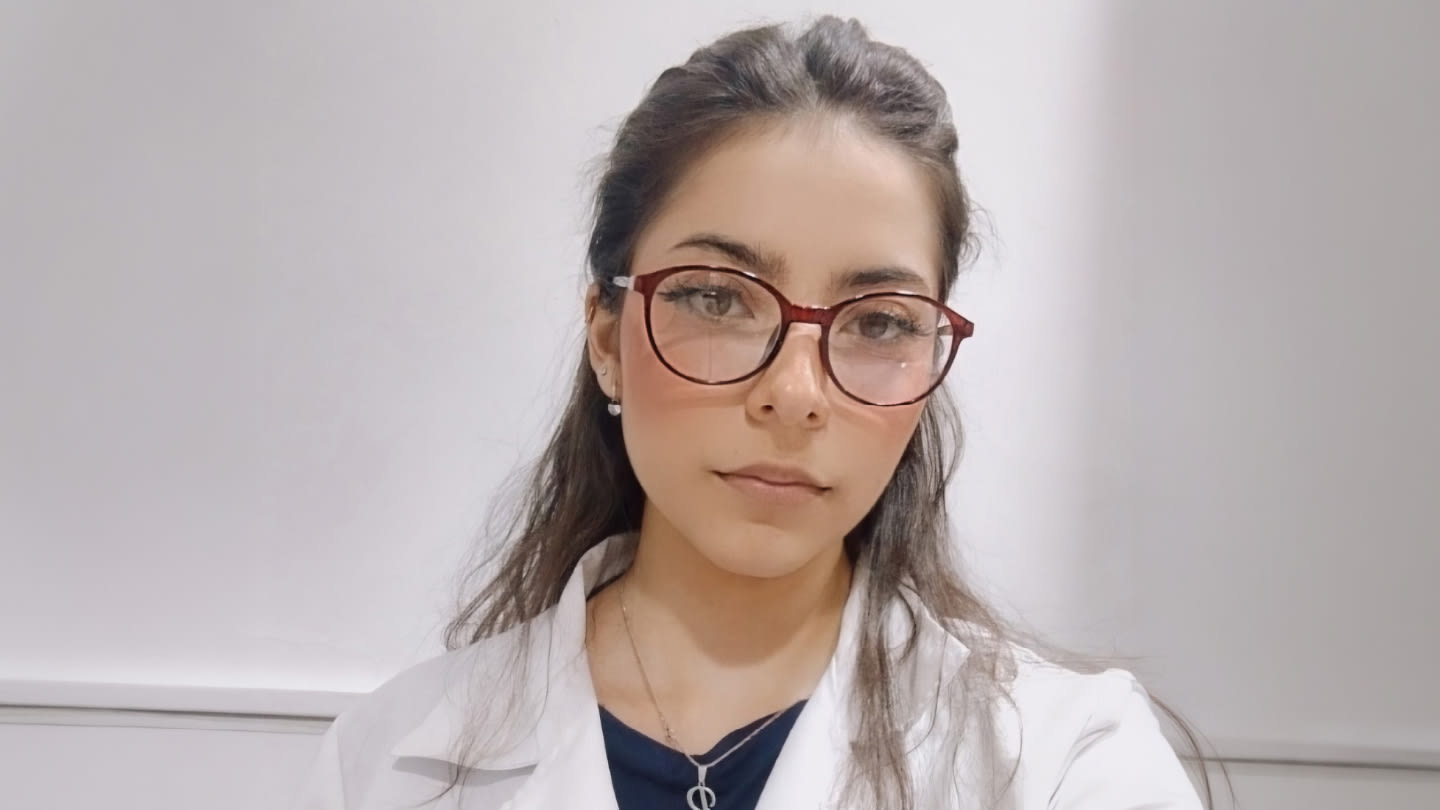
Agar art has offered a new perspective on developing educational projects in pathology, microbiology, and laboratory science. By transforming complex concepts into visual representations, it enhances understanding, supports learning, and makes scientific communication more engaging and accessible.
Fiorella Páez and Camila Robayo
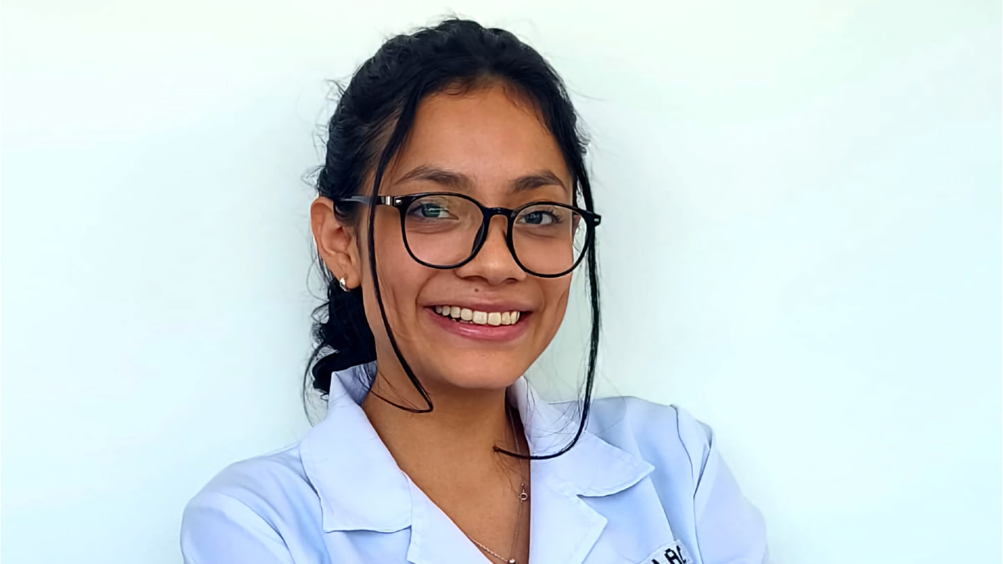
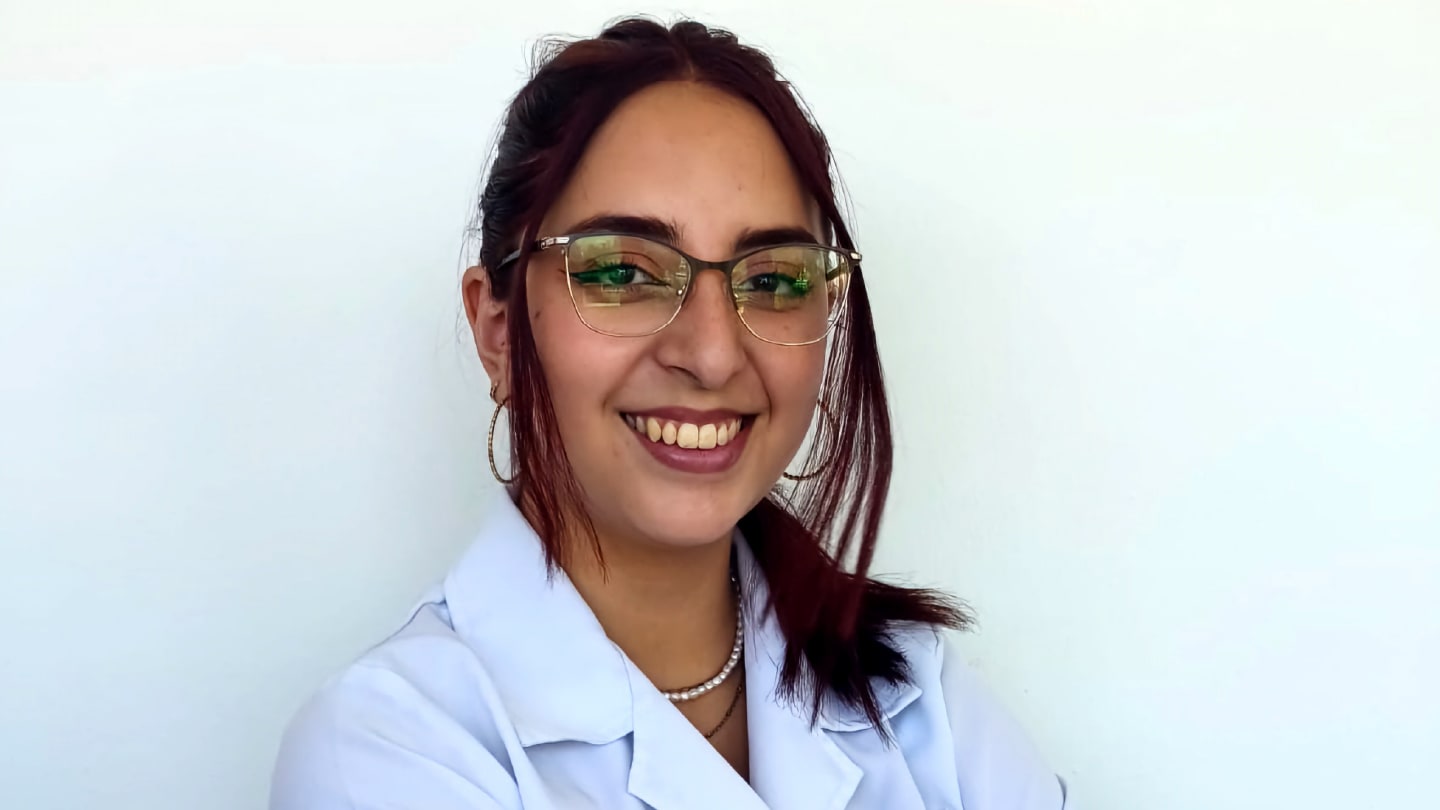

These projects allowed us to learn about different microorganisms, including their morphology and specific characteristics, while integrating the knowledge we had acquired throughout the semester.
Participating in these projects made the learning process more interactive and engaging. Creating the artwork helped us understand and remember the microorganisms’ distinguishing features, growth patterns, and colors.
The experience transformed learning from a passive process into an active one, reinforcing concepts in a way that traditional lectures and classes could not.
Alejandro Terán
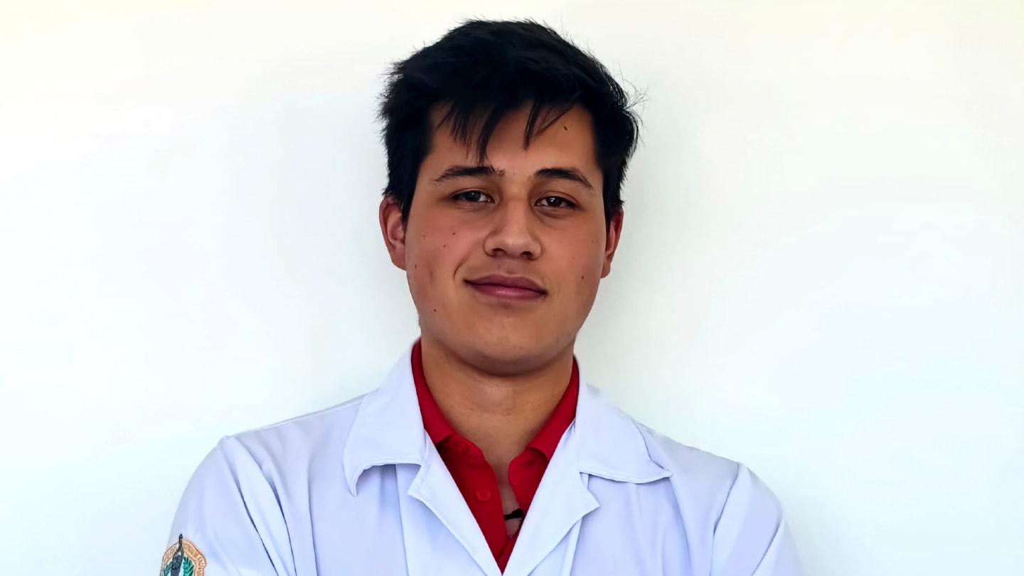
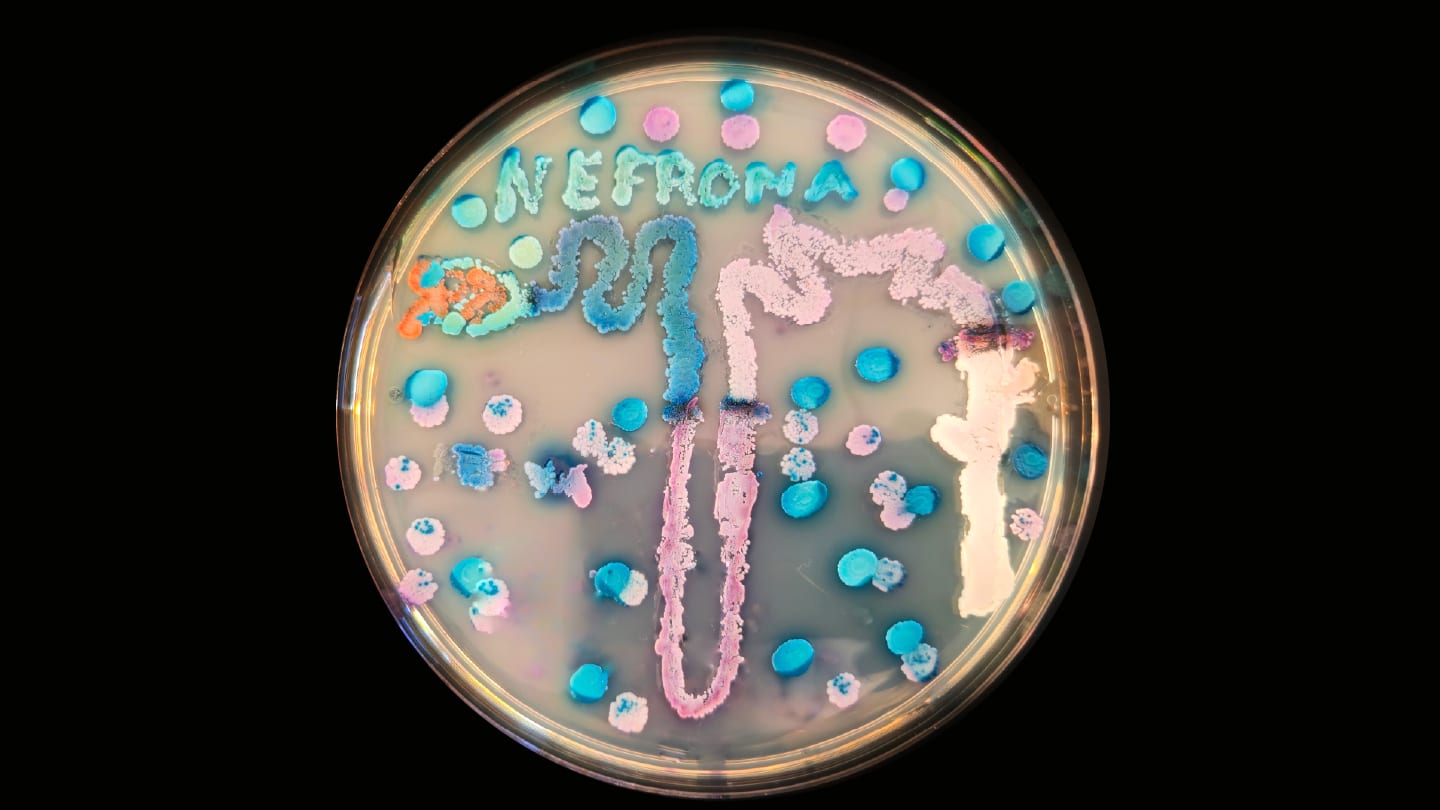
Physiology is one of the aspects of my profession that I enjoy most. Agar art helps me identify the different components of an organ or structure and understand the function of each one. It also demonstrates how an abnormality in any of these components may contribute to disease and how recognizing such changes can support early diagnosis.
The activity also helps me distinguish the characteristic colors of different yeast species. In “The Art of Filtration,” for example, I learned about the parts of the nephron and the importance of filtration and excretion.
Ariana Bastidas
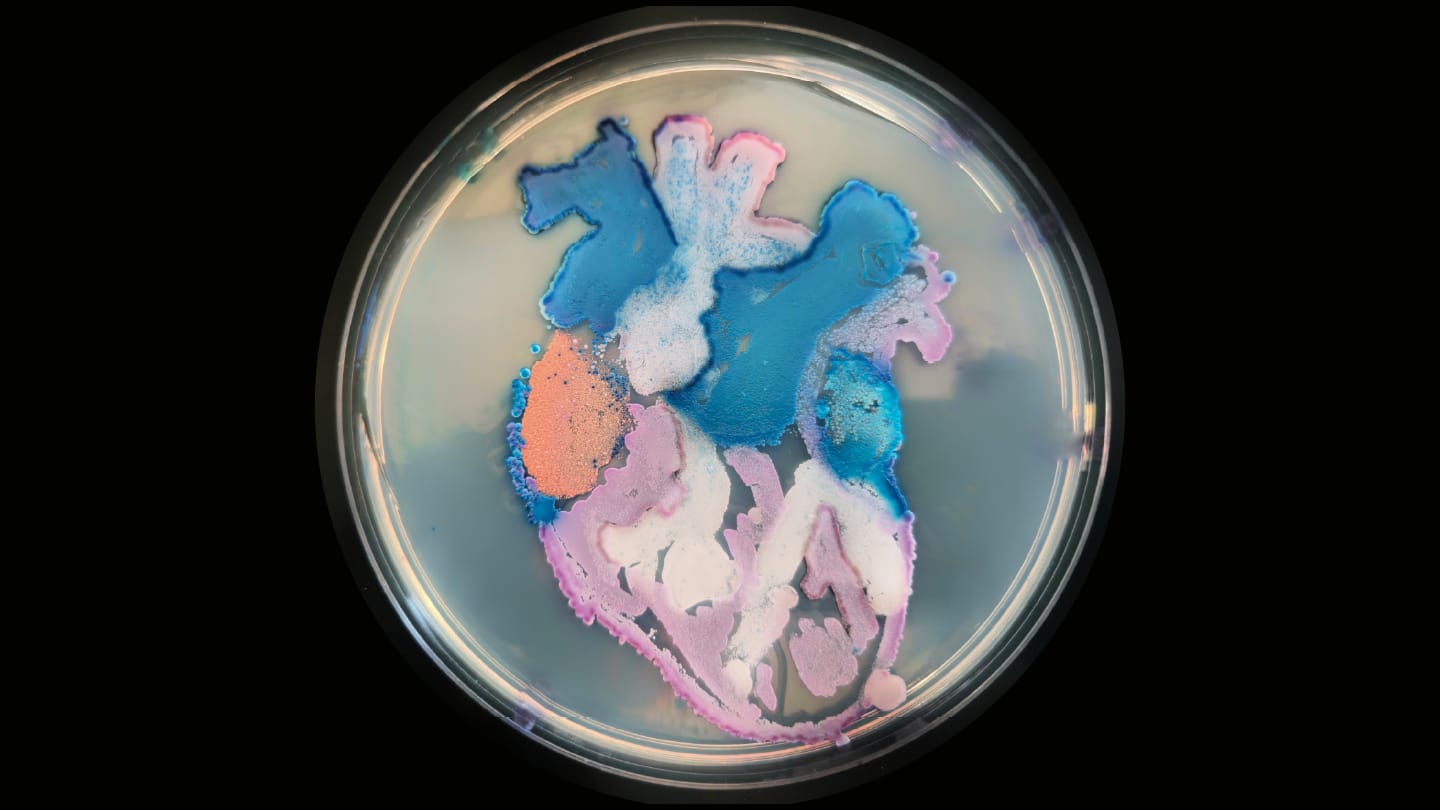
Creating my agar art project deepened my practical understanding of microbiology by demonstrating how yeast species produce distinct colors on chromogenic media. Using different strains to create an image of a heart made species differentiation and identification feel tangible rather than purely theoretical. The project also connected laboratory isolation techniques with the diagnosis of specific infectious diseases.
How have students benefited from creative teaching methods, whether through engagement, confidence, or understanding of the subject matter?
The response has been overwhelmingly positive. Students become more engaged when they create actively rather than memorize information passively. Seeing their initial sketches transformed into living microbial artwork builds confidence and inspires curiosity. The experience also demonstrates that learning laboratory medicine can be both scientifically rigorous and highly creative.
Do you think creative approaches such as #PathArt will play a bigger role in the future of pathology and diagnostics education?
Absolutely. I believe creative approaches will play an increasingly important role in pathology and laboratory medicine education. By combining scientific knowledge with clinical reasoning, visual learning, and creativity, these methods help students connect classroom concepts with real clinical practice. As diagnostic technologies evolve, strong observational and interpretive skills will remain essential, and creative teaching methods can help students develop these abilities.
Newsletters
Receive the latest pathologist news, personalities, education, and career development – weekly to your inbox.