For many years, rapid on-site evaluation (ROSE) at Washington University School of Medicine (WashU) looked much like it does in hospitals everywhere: cytopathologists or cytotechnologists shuttling from one procedure room to another, waiting for slides to be prepared, reviewing them under a microscope, then rushing back to another site across a sprawling campus. The process was functional, but it became more difficult to manage and increasingly unsustainable to maintain as procedure volumes and diagnostic complexity increased.
"Some days, I spent nearly an hour just walking between sites for on-site evaluations," recalls Hannah Krigman, Professor of Pathology and Immunology at WashU. "That's an entire hour of lost diagnostic time."
The challenge isn't unique to WashU. Rising cancer incidence, growing demand for molecular testing, and a persistent pathologist shortage, with only 65 per million people in the US, have been straining cytology services nationwide. WashU needed a solution that could extend expert coverage across its expanding network without compromising speed, quality, or patient experience.
Finding the right technology
When WashU's cytopathology team began exploring digital solutions, they had specific requirements. The technology needed to produce real-time images from procedure rooms, match traditional microscopy quality, and integrate seamlessly into high-pressure ROSE workflows. Large, centralized scanning systems requiring slide transport wouldn't work.
"We wanted something that felt as natural as sitting at a microscope, but without requiring physical presence," says Suzanne Crumley, Associate Professor of Pathology and Immunology at WashU. "The form factor mattered; in a procedure room, space is limited, and workflow is everything."
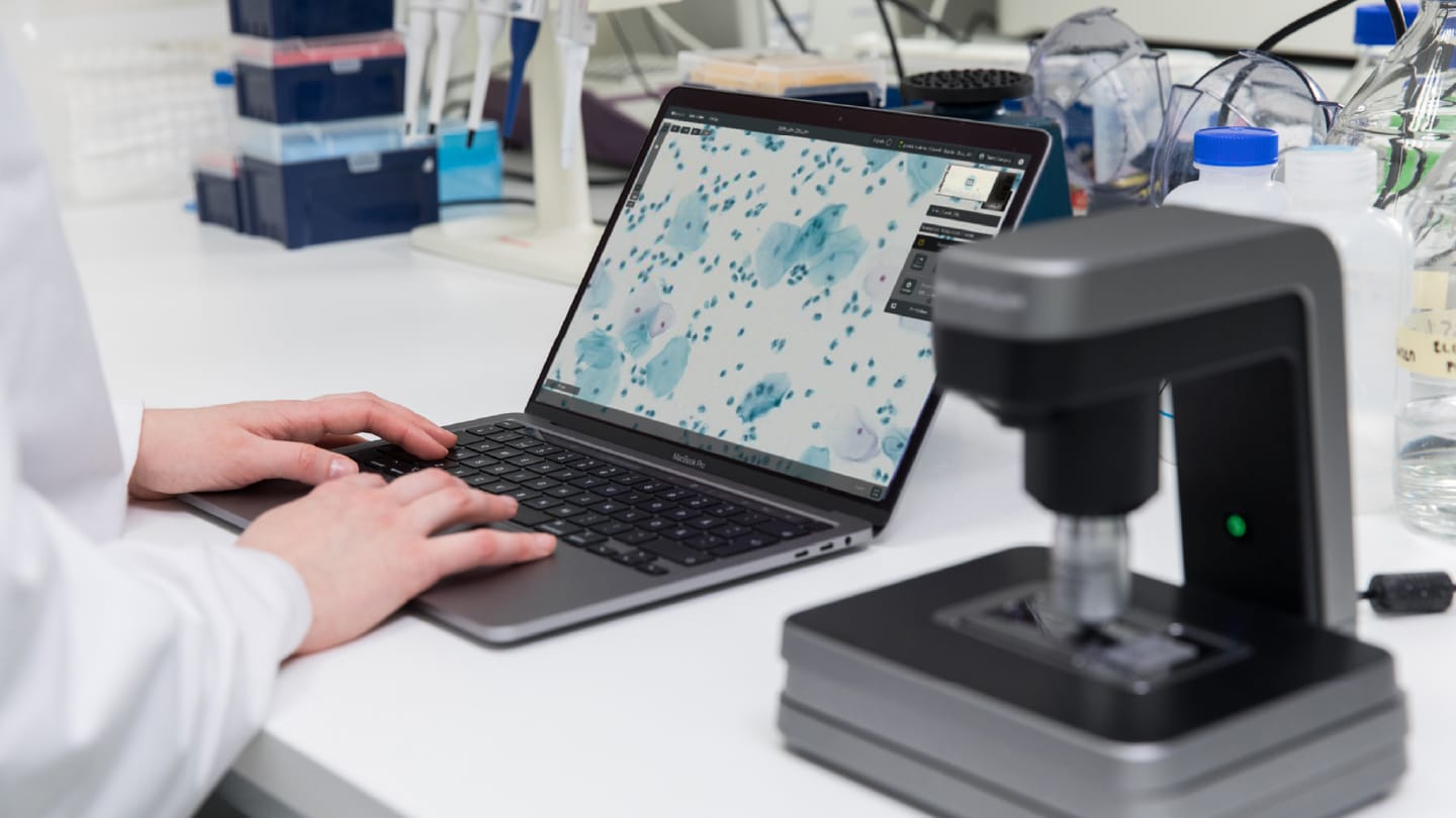
The collaboration with Grundium, a specialist in digital imaging for pathology, offered the required approach: compact scanners small enough to position directly in procedure rooms. Rather than transporting slides to a scanning facility, cytotechnologists could scan them immediately after preparation, with high-resolution images available for remote review within minutes.
The browser-based system eliminated complex software installations. Pathologists could access images through secure cloud transmission, panning, and zooming just as they would at a microscope. Crucially, the "live view" capability enabled real-time viewing of specific slide areas without waiting for complete scans, essential for the time-sensitive nature of ROSE.
Expanding coverage while maintaining quality
By 2023, the program encompassed six locations, including two Illinois hospitals with insufficient case volumes to justify dedicated permanent on-site cytopathology staff. For these remote sites, WashU deployed the Ocus 40 scanner, which captures images at 40x magnification, standard resolution for diagnostic cytology.
The workflow operates with minimal complexity. Cytotechnologists prepare and scan slides at remote sites, while pathologists review images from a central location. Communication flows via phone or secure messaging. If a specimen proves inadequate, the pathologist notifies the clinician immediately, while the patient remains in the room, enabling the potential collection of additional material.
Before clinical implementation, WashU conducted rigorous validation comparing digital evaluation against traditional on-site microscopy. The process revealed an unexpected benefit: improved slide preparation consistency across sites. "The digital archive created a permanent record of each case," Krigman notes. "In a network our size, this helped us coordinate how slides were prepared, scanned, and reviewed."
Similar validation at other institutions has demonstrated robust concordance. Intermountain Health reported 94 of 96 reads in full agreement when comparing digital frozen sections to glass slides.
Quantifiable impact
The efficiency gains proved substantial. By eliminating travel between sites and reducing waiting periods, WashU effectively reclaimed an additional workday of diagnostic time per pathologist each week, translating into capacity for more procedures without increased staffing.
Clinical benefits extended beyond logistics. Real-time adequacy assessment prevents inadequate samples that would necessitate repeat biopsies, sparing patients additional procedures while ensuring sufficient tissue for increasingly critical molecular testing. Preliminary diagnoses often also arrive faster than traditional workflows, as clinicians no longer wait for a pathologist's physical return.
"With digital ROSE, we confirm specimen adequacy immediately, even from across campus," Crumley explains. "That protects patients from repeat biopsies and ensures we have material for molecular workups."
Implementation lessons
Success required more than installing scanners. WashU worked closely with the vendor to streamline the entire workflow: slide preparation techniques, scanning protocols, quality control procedures, and communication pathways between remote sites and pathologists.
"The technology works, but success depends on workflow integration," Crumley comments. For WashU, this meant training cytotechnologists to prepare slides ready for digital imaging, establishing clear communication protocols, and ensuring adequate network bandwidth and compliance with security requirements.
Change management proved critical. Initial concerns from some pathologists dissolved once they experienced the image quality and workflow efficiency firsthand. The compact scanner design facilitated adoption, unlike large systems requiring dedicated space, the small scanners could be deployed quickly for phased rollout.
Expanding access to expertise
The benefits of digital ROSE extend beyond operational efficiency. Many hospitals struggle to maintain specialized cytopathology coverage across multiple sites, while rural centers often lack expertise entirely. WashU's model demonstrates how point-of-care scanning enables expert support for facilities that otherwise couldn't access it.
"With a secure digital link, multiple satellite centers can be supported without sending a specialist on the road. This is how pathology becomes more equitable," says Todd Vanden Branden, Grundium's Senior Director of Marketing and Field Applications.
With approximately 1,000 pathologist positions unfilled in the US, but only 450 new pathologists entering the field annually, technology-enabled service delivery models may be essential for maintaining access to specialized diagnostics, particularly in underserved regions.
Looking ahead
With the foundation of infrastructure established, WashU envisions expanding beyond ROSE to frozen sections, multidisciplinary tumor boards, remote second opinions, and eventually AI-assisted interpretation. This follows success at Intermountain Health, which deployed similar technology across eight sites in Utah to support telepathology for frozen section support.
"Digital ROSE allows us to bring subspecialty-level expertise into every case. As diagnostic complexity increases, that capability becomes increasingly important," says Crumley.
The collaboration between WashU and Grundium demonstrates how compact, point-of-care scanning technology can address real-world challenges in pathology. For clinicians performing biopsies, pathologists interpreting them, and patients awaiting answers, this represents a fundamental reimagining of how specialized services can be delivered: faster, more consistently, more accessibly, and better aligned with workforce realities facing modern medicine.
As institutions worldwide confront similar pressures, the WashU experience offers a blueprint. With thoughtful technology selection, robust validation, and strategic implementation, digital telepathology can transform critical bottlenecks into opportunities for improved patient care.
Newsletters
Receive the latest pathologist news, personalities, education, and career development – weekly to your inbox.


