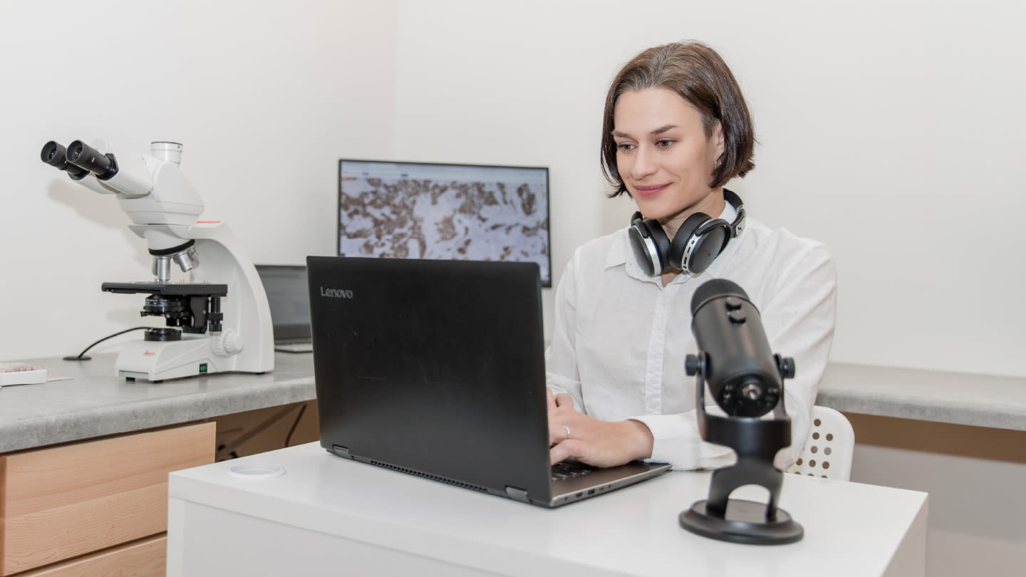
From veterinary pathology to podcasts, Aleksandra Zuraw has been a central figure in starting conversations about digital pathology. Following her keynote presentation at DP&AI: Europe in London December 2025, she joined us to share her motivations, learnings, and advice.
Meet Aleksandra Zuraw
I originally thought I would become a horse veterinarian. After three years working as a general practitioner veterinarian, I knew I wanted to pursue a PhD and continue learning, but in Poland there were limited opportunities for internationally recognized specialization.
I began exploring the European Board of Veterinary Specializations and reviewed the full list of options. Two specialties stood out to me: pathology and equine medicine. I looked for institutions that offered both and eventually identified an opportunity in Berlin, Germany, which was geographically close and offered a scholarship. The plan was to spend six weeks in a pathology department and six weeks in a horse clinic.
I started in the pathology department, and about three weeks in, I called the horse clinic and said, “I’m not coming – I’m staying here.” I realized that pathology could truly be a career. In Poland, most veterinary pathologists worked exclusively in academia, which I did not want. Even though I enjoyed the subject, that pathway felt too limiting.
My path into digital pathology happened almost by accident. While finishing my residency in Berlin and completing my PhD project, I was given a study that required quantifying immunohistochemistry staining. Visual assessment was extremely difficult, so I began taking digital images and – quite primitively – annotating them in Microsoft Paint. That was obviously far removed from the digital pathology tools we use today, but it was my entry point.
Through that process, I realized that digital images allowed for quantification, reproducibility, and objective analysis. That small project gave me a competitive edge during job interviews and ultimately helped me secure a position at an image analysis company after completing my PhD. From there, I never looked back.
Veterinary pathology involves a wide range of species and tissue types. How has this diversity shaped your adoption of digital tools?
At first glance, digital pathology in veterinary medicine may seem highly complex, but it is more manageable than it appears – especially when focusing on mammals. While there are many mammalian species, their basic tissue architecture is largely similar.
Visually, both MD and DVM pathologists can usually recognize the same tissues. The key differences lie in diagnostic interpretation. Veterinary pathologists encounter diseases that may not exist in humans, may be named differently, or have different prognoses. So although tissues may look alike, the diagnostic context can vary.
This becomes particularly important in digital pathology and AI, which are highly sensitive to domain-specific data. Differences can arise from scanners or institutions, but species variation is especially significant. An AI model trained on one species does not automatically work for another.
For example, a model developed for mouse tissue will not reliably perform on rat or canine tissue without additional training. The structures are similar, but not identical enough for AI to generalize on its own.
As a result, even with advanced AI, digital pathology tools still require species- and tissue-specific development, along with careful validation to ensure accuracy and reliability.
In what ways can veterinary pathology contribute to the development and validation of computational pathology tools?
From an industry perspective, I focus on applications that are both feasible and high-impact. The key question is always: What is the lowest-hanging fruit that delivers the greatest value? That means tools that are practical to build, easy to deploy, and clinically meaningful.
In toxicologic and diagnostic pathology, this often points to tasks that are visually straightforward and high-volume. For example, mast cell tumor detection in dogs – already supported by commercial tools – is well suited to computer vision because the features are clear and clinically relevant.
The next consideration is impact. If a diagnosis does not meaningfully influence treatment decisions, the return on developing an automated tool is limited. High-value applications are those where results directly affect clinical management.
Once a use case is chosen, development follows a familiar path: defining the problem, assessing the market, building the software, and deploying it in practice. Even when an application is specific to veterinary pathology, the development process itself provides lessons that others can reuse.
Veterinary pathology also moves faster because it faces fewer regulatory barriers than human medicine. This makes it an effective testing ground, allowing teams to iterate quickly, generate evidence, and share insights that can later inform digital pathology development across disciplines.
The Digital Pathology Place podcast has grown into a global learning hub. What motivated you to create it?
The podcast opens with the phrase “bridging the gap between pathology and computer science,” which reflects a challenge I experienced firsthand in my first industry role. I came in as the tissue expert, working alongside computer vision specialists. It took several months for us to develop a shared knowledge base – where they understood enough pathology and I understood enough computer vision – to collaborate effectively. That alignment didn’t happen automatically; it took real effort, but once it did, it enabled meaningful projects and rapid progress.
That experience made it clear to me that this gap was not unique to one company. If it existed within an organization dedicated to digital pathology, it likely existed everywhere – especially outside highly specialized environments. When I later moved into a more traditional toxicologic pathology role, I wasn’t sure how directly I would continue working with image analysis, but I knew I wanted to stay connected to the digital pathology space.
That motivation led me to start the blog and the podcast. Over time, the focus has broadened. With AI now firmly in the picture, the challenge is no longer just about pathology and computer science, but about bridging medicine and life sciences with computational disciplines more broadly.
Through your podcast conversations with leaders across clinical, veterinary, and computational pathology, what recurring themes or misconceptions have you observed about digital pathology’s role in diagnostics?
One misconception I often address is the idea that you need extensive infrastructure to start working with digital pathology. People assume you must have a whole-slide scanner, but that simply isn’t true. You can begin with something much simpler.
In the veterinary world, for example, a colleague of mine built a diagnostic clinical pathology business by capturing microscope images with a smartphone and sharing them through an app. Digital images – at their most basic level – are the foundation of her practice. That really shifted my perspective, because we often think of digital pathology only in terms of scanners and whole-slide images, when in reality those are just one part of the ecosystem.
The question then becomes how else we can leverage digital data. With tools like large language models now available, there are new ways to extract value from image data, even when it’s captured in low-resource settings. This opens the door to greater accessibility, second-opinion workflows, and broader adoption – especially in areas with fewer regulatory constraints, such as veterinary medicine.
Another common challenge is not technology, but change management. Everyone is at a different stage in their digital pathology journey. Some are just starting, others are well underway, and there will always be early adopters alongside more skeptical colleagues. Navigating that cultural shift remains one of the biggest hurdles.
The key lesson from many digital pathology pioneers is simple: start with what you have. Technology will always evolve, and today’s cutting-edge tools will eventually be replaced. Progress comes from using available resources, not waiting for the perfect setup.
What skills do today’s digital pathology “trailblazers” need – beyond technical curiosity – to effectively lead transformative diagnostic projects?
We are in an exciting moment where the new generation of pathologists is increasingly tech savvy. I consider myself comfortable with technology, but when I speak with colleagues who are ten years younger, it’s clear they are truly digitally native.
That said, technical comfort alone is not enough. What we need is a strong commitment to continuous education. This was a key message in my talk. The field is evolving so quickly that learning can no longer be a one-time event. It is not a matter of taking a single course and being “certified.” New tools, models, and approaches emerge constantly, so staying current requires ongoing effort.
It also requires an open mindset. In digital pathology, no one person can be the expert in everything. Pathologists bring deep domain knowledge, but we increasingly work within multidisciplinary teams that include data scientists, engineers, and other specialists. This is especially true in the pharmaceutical industry.
To work effectively in this environment, you need to develop a shared knowledge base – enough understanding of each other’s roles to collaborate productively. That was something I had to learn early on: knowing who the experts are, what they contribute, and how your own expertise fits into the larger ecosystem.
How important are community building and peer-to-peer leadership for safe, successful digital pathology adoption?
In a fast-moving field like this, the pace of technological change can be intimidating. It can be hard to approach thought leaders or vendors if you’re not yet sure what you need or even what questions to ask. That’s why finding someone who is just one step ahead on the same journey can make such a difference. It’s less intimidating and gives you access to practical, behind-the-scenes insights – especially about mistakes that rarely get published, since negative results and missteps are often left out of the literature.
Building relationships with people at a similar stage helps you avoid common pitfalls and move forward with more confidence. That idea is central to what I wanted to create with the digital pathology trailblazer community.
I also believe strongly in being open and approachable. During my talk, for example, my smart glasses started speaking unexpectedly, and I had to stop and turn them off. Moments like that show vulnerability, and they make it easier for others to reach out. For me, it’s important that education in digital pathology remains accessible and welcoming, no matter where someone is in their journey.
What teaching strategies have you found most effective for helping diagnosticians build confidence with digital and computational tools?
One of the fastest ways to learn in digital pathology and AI is to work toward a concrete goal. If you can be part of a project – developing an algorithm, using software to solve a specific problem, or improving a workflow – you will learn much more quickly than by simply watching demos or exploring tools in the abstract.
The key is to learn with purpose. That goal might be something small, such as quantifying positive cells in an image, or something broader, like streamlining a histology workflow. Having a clear objective makes the learning process more focused and less overwhelming – you learn what you need as you go.
Not everyone can immediately join a digital pathology project, and that’s okay. The next best step is committing to consistent learning. Choose one reliable source and engage with it regularly – whether that’s attending live streams, following a trusted voice in the field, setting up PubMed alerts, or reading publications like The Pathologist. You don’t need to do everything; just pick what resonates and stick with it.
Over time, this approach compounds. A year from now, you’ll notice that you understand concepts you weren’t even aware of before. I see this myself when I revisit podcast conversations – each time I listen, I pick up new insights, simply because my knowledge has grown.
Which developments in digital and computational pathology do you think are having the greatest practical impact on laboratories right now?
Two developments have really surged: agentic AI and end-to-end digital pathology workflows.
Agentic AI refers to AI systems that coordinate other AI tools. Instead of performing a single task – such as detecting a specific cell type – it orchestrates multiple systems. For example, one AI might retrieve an image, another performs image analysis, and a large language model interprets the results in a format a pathologist can easily review. The value lies in automation and coordination, not just analysis.
This has enormous potential for pathology workflows. In both veterinary and human pathology, experts spend significant time navigating fragmented systems, clicking through databases, and managing inefficiencies. Agentic AI can reduce that friction by bringing information together and presenting it in a usable way.
The second major trend is the move toward end-to-end workflows. We are now at a point where many digital pathology tools – both hardware and software – have regulatory approval and are ready for clinical use. That means early adopters have more choices than ever, but it also means no single tool will stand alone.
Interoperability is therefore critical. The most effective solutions are those where scanners, image management systems, and other tools already work well together. Choosing vendors with proven, integrated workflows – from staining through sign-out – can dramatically reduce complexity and help laboratories become digital-ready much faster.
Finally, what advice would you offer to the next generation of pathologists and digital trailblazers?
The best advice I can give is continuous learning. I don’t know whether this is generational, but there’s often an expectation that by the end of training, you should have everything figured out and commit fully to one path. In reality, careers evolve, and pivoting is normal.
The best way to decide where to pivot is to stay engaged with what genuinely interests you. If that interest is digital pathology, choose one avenue for ongoing learning and follow it over time. That process will naturally guide you toward your next step.
Newsletters
Receive the latest pathologist news, personalities, education, and career development – weekly to your inbox.


