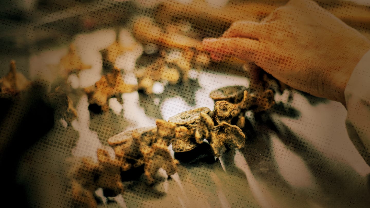
Thanks to their mineralized structure, bones are the only organ capable of surviving for thousands of years. This unique property makes them an extraordinary archive of information about our ancestors – the biological time capsules of the human race. From morphological observations to advanced molecular analyses, studying ancient bones can reveal how people lived, what they ate, and even what diseases they faced.
Take osteoporosis, for instance – a condition characterized by the loss of bone density that is typically associated with modern aging, especially in postmenopausal women. We often consider it a disease of the 20th or 21st century, but history tells a different story.
Paleopathological research indicates that osteoporosis has existed for a long time, affecting Egyptians, Romans, and Vikings alike. A particularly compelling study by Zaki and colleagues examined 74 skeletons from the Giza Necropolis, dating back to Egypt’s Old Kingdom (2687–2191 BCE). They found that bone loss varied according to social status: the fewest signs of osteoporosis were in upper-class men, followed by male laborers and women. The most severe cases were observed in upper-class women – those who enjoyed rich diets but led sedentary lives. This pattern closely mirrors what we see today: poor nutrition and lack of physical activity remain key risk factors for osteoporosis. Once again, lifestyle matters across all ages and eras.

Another fascinating disorder revealed through ancient evidence is Paget’s disease, a metabolic bone condition caused by the excessive activity of bone cells. It leads to abnormally large and deformed bones, often affecting the skull and face.
A possible depiction of this disease can be found in the National Gallery in London. There, a painting by Quentin Matsys from around 1513 – The Ugly Duchess (also known as Grotesque Old Woman) – portrays a woman with striking facial deformities. Though often dismissed as caricature or satire, medical experts have noted that her features closely resemble those of modern patients with Paget’s disease. Michael Baum proposed this theory in a 1989 article in the British Medical Journal, suggesting that the woman in the painting may have suffered from the condition.
Paget’s disease was first described in 1877 by Sir James Paget, who believed it to be a newly discovered illness of his time. Yet, ironically, evidence of it may have been captured in art centuries earlier.
The story stretches even further back into history. In Norse legend, the Viking warrior Egil, son of Skalla-Grim, was described as terrifying in appearance, with exaggerated features and bone deformities. Many believe Egil may also have had Paget’s disease, a powerful reminder that even mythic figures might have been shaped by real medical conditions.
Going back 4,700 years to ancient Egypt, we also find King Sanakht, a pharaoh of the Third Dynasty who was described as a “giant” and even revered as a god. But was he truly divine, or was his extraordinary height the result of gigantism, a condition caused by the excessive secretion of growth hormone? In 2017, researchers examined what are believed to be his remains and found that he was significantly taller than others from the same period. If this diagnosis is correct, King Sanakht would be the oldest known case of gigantism in history.
I could talk for hours about benign and malignant bone tumors, but even a brief glimpse into their history is fascinating. It turns out that bone tumors like osteosarcoma, which we often think of as modern diagnoses, have been around far longer than we might imagine. Signs of osteosarcoma have been discovered in ancient Egyptian skulls as well as in the fossilized remains of a human relative dating back 1.7 million years. This astonishing finding shows that cancer is not a disease of our industrial age; it’s part of our deep evolutionary past.
And our discoveries of ancient disease don’t stop at humans. Evidence of benign bone tumors, such as hemangiomas and osteoblastomas, has been identified in the fossilized bones of ancient dinosaurs. These findings suggest that even Earth’s most colossal creatures were not immune to the diseases we face today.
But perhaps the most captivating story comes from a skeleton discovered in 2003 in La Noria, Chile – a specimen known as Ata. With her elongated skull and small, narrow body, Ata’s appearance sparked wild theories, including claims that she was an extraterrestrial being. Her skeletal deformities were so unusual that they challenged the limits of what was known about human anatomy.
Yet, science offered a more grounded – though no less remarkable – explanation. DNA analysis revealed that Ata was a stillborn human girl who had multiple skeletal abnormalities caused by mutations in at least seven genes. These mutations affected bone development and cranial structure, resulting in her distinctive, alien-like appearance. What looked like science fiction was, in fact, a profound example of how genetics and rare disorders can shape the human form in unexpected ways.
These stories, preserved in both skeletal remains and art, reveal an important truth: many of the diseases that we consider modern have existed for thousands of years. Whether found in the ruins of ancient tombs or immortalized in paintings and legends, these conditions offer remarkable insights into the health, lives, and humanity of those who came before us.
In the end, ancient bones don’t just tell us how people died – they tell us how they lived.


